Use our HearthHealth app
During exercise, healthy coronary arteries dilate (stretch and develop a more open channel) more than an artery that has a blockage. This unequal dilation causes more blood to be delivered to heart muscle supplied by the normal artery. In contrast, narrowed arteries end up supplying reduced flow to it’s area of distribution. This difference in blood flow can be detected by a “perfusion tracer” (a nuclear isotope that travels to heart muscle with blood flow) injected intravenously.
The perfusion tracer is extracted by the heart muscle in proportion to the flow of blood. The amount of tracer uptake helps differentiate normal muscle (which receives more of the tracer) from the reduced uptake demonstrated by muscle that is supplied by a narrowed coronary artery. In other words, areas of the heart that have adequate blood flow quickly picks up the tracer material. In contrast, muscle with reduced blood flow
pick up the tracer slowly or not at all.
Analysis of the images of the heart (taken by a scanning camera) can help identify the location, severity and extent of reduced blood flow to the heart. The reduced blood flow is known as ischaemia.
The test is actually divided into three parts: Imaging at rest, a treadmill stress test, and imaging after exercise. There are two common types of isotope used: thallium and technetium. Some laboratories use a “dual isotope” technique, where thallium is used for the resting images and technetium is used for the stress pictures. Depending upon the isotope and protocol for the laboratory, resting images may be obtained either before stress or two to four hours after stress.
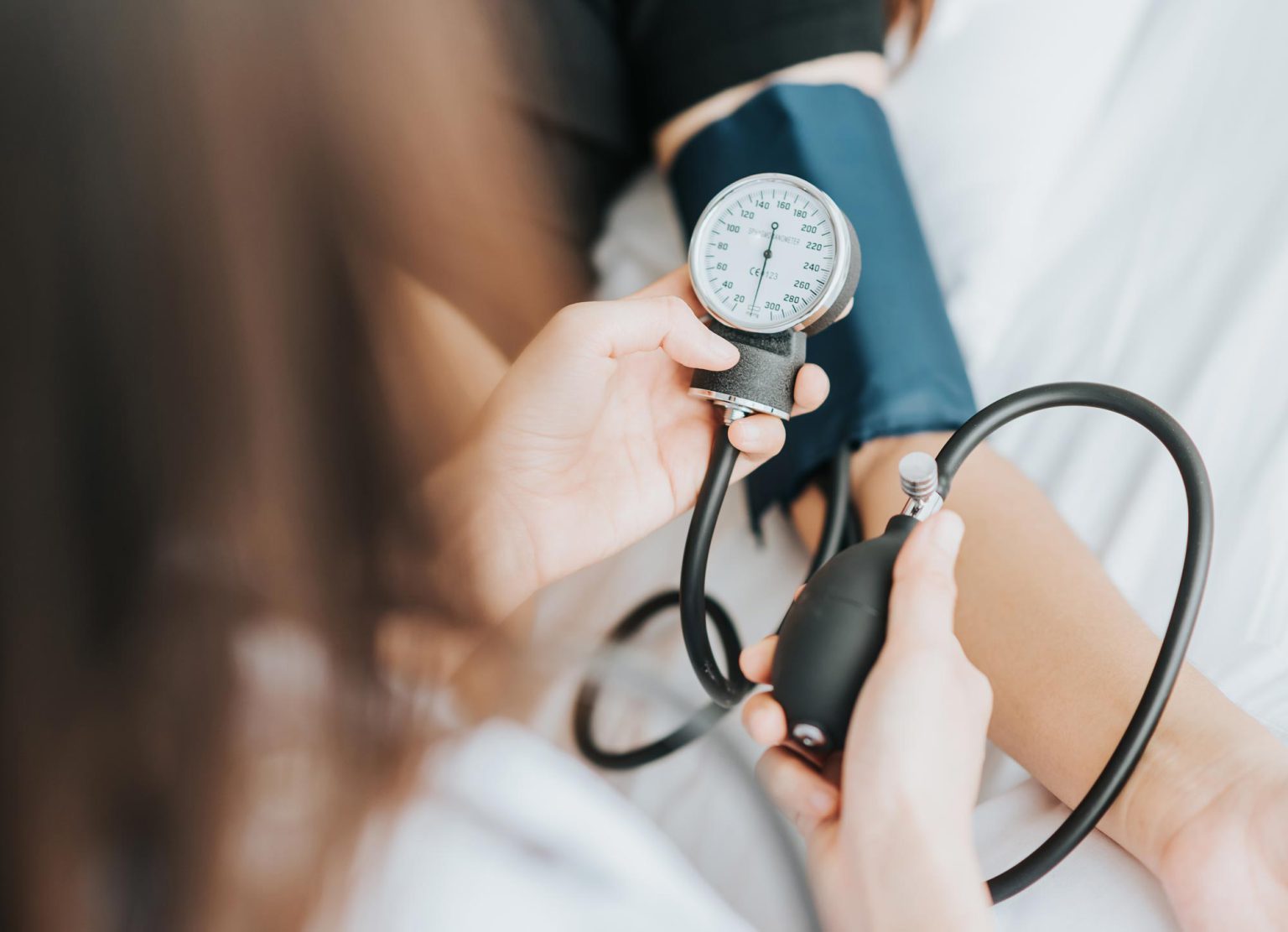
Your heart rate and blood pressure will be monitored throughout the test, and the ECG is displayed continuously on a monitor and printed every minute. About one minute prior to termination of exercise, the perfusion tracer or isotope is injected into your vein. After a brief wait (to allow the tracer to be taken up by the heart muscle) you will be placed under a scanning camera. The scanning camera rotates around the patient’s chest, stopping to take individual pictures. You will need to lay flat and still during the scanning period which takes approximately 11 to 20 minutes, depending upon the type of scanning camera. The pictures or images are fed into a computer, which reconstructs them as “slices” of a three dimensional heart.
You are exposed to a very small amount of radiation and the risk is minimal, if any. The risk of the stress portion of the test is very small and similar to what you would expect from any strenuous form of exercise (jogging in your neighbourhood or running up a flight of stairs). Experienced medical staff are in attendance to manage rare complications like sustained abnormal heart rhythm, unrelieved chest pain or even a heart attack. The risks are very small – if you have any concerns please discuss them with your Cardiologist prior to the procedure.
If you are able to achieve your target heart rate (85% of maximum) and good quality images are obtained, a myocardial perfusion scan is capable of diagnosing important disease in approximately 85% of patients with coronary artery disease. Approximately 10% of patients may have a “false-positive” test (when the result is falsely abnormal in a patient without coronary artery disease). Technical problems can occur if you are markedly overweight. Women may have an abnormality in the front portion of the heart because of overlying breast tissue. Some men may demonstrate an inferior wall abnormality because of a prominent diaphragm (muscular partition that separates the chest cavity from the abdomen). Patients who have a left bundle branch block on their ECG may also have a false abnormal test.
The doctor performing the test can give you a preliminary report about your test. However, the official result from the isotope scans may take a few days to complete as the images need to be analysed in detail. The results may influence your doctor’s decision to change your treatment or recommend additional testing such as cardiac catheterisation.